Cases Archives - Page 2 of 2 - Veterinary Specialists Aotearoa | Veterinary Specialist and Animal Referral Services in NZ with 3 specialist animal hospitals in Auckland and Christchurch

Cases

LUCKY TESTS HER LUCK

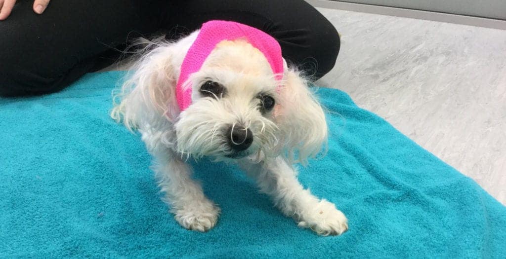
Lucky, a feisty little white female Maltese, had a pretty bad day on Thursday the 9th of June.  She was hanging out at home being inquisitive and guarding the home base when she tipped her luck too far and fell out of the window. Dropping two floors onto a hard surface causing spinal cord damage and immediate paralysis from the neck down. Vicky, her Mum, rushed her to their vet Dr Deborah Jones at the Kohimarama Veterinary Clinic for emergency care. A decision was made to refer her to VSA for surgical assessment. Lucky was seen by Dr Richard Jerram who localised the spinal damage to C1-5 (cervical 1 through to 5) and recommended a CT of that area of the spine. Lucky was in a lot of pain but could feel and move her legs and had normal mental awareness. The CT was read by Dr Angela Hartmann and showed “a comminuted fracture associated with the occipital bone and basioccipital bone.  This has likely resulted in atlantooccipital instability given that both occipital condyles are involved.  There is at least one large segmental fracture associated with the right caudal portion of the occiput.  No intracalvarial hemorrhage is noted.” This means that Lucky had probably landed on her head and the spinal vertebrae had pistoned into the back of her skull fracturing the bone (occipital bone) and compressing the brainstem/cranial cervical spinal cord.  Note the bone fragment displaced cranially and compressing the brain stem. Note the fractured occipital condyle on the left and the right foramen fracture. After a detailed discussion with Vicky it was decided to take Lucky to surgery with the goal of decompression of the brain/spinal cord and stabilisation of the atlanto-occipital joint.  At surgery (Drs Jerram and Walker) we identified a large displaced piece of bone compressing the spinal cord. This was removed and the base of the brain (cerebellum) was decompressed by careful removal of some more of the occipital bone.  Once the decompression was achieved Lucky’s head was then positioned in a normal standing position and stabilisation between the skull and the dorsal aspect of the C1 vertebra was performed by creating a custom device using titanium mesh and self tapping screws.  Five screws were placed in the dorsal arch of the C1 vertebra and six screws were placed in the transverse sinus region of the skull.  Following completion of this procedure, the skull and the C1 vertebra moved together (see radiograph below).   Lucky was placed in a neck splint and immediately showed improvement in the post-op period – marked reduction in pain when manipulated and stronger motor movement.  Lucky could ambulate with assistance within one week and was walking strongly within a month.  Lucky is still wearing the neck brace at 6 weeks post-op because she is trying to run around and is nearly normal neurologically. This case demonstrates the huge advantage to having access to an in-hospital CT and without that equipment this life-saving surgery would not have been possible. Thank you to Vicky for letting us tell Lucky’s story. DR ALEX WALKER Director & Registered Specialist Small Animal Surgery  alex.walker@vsnz.co.nz Alastair graduated from Massey University with a Bachelor of Veterinary Science in 2003, and worked in mixed animal practice in Ashburton for 18… READ MORE… Share on facebook Facebook Share on twitter Twitter Share on linkedin LinkedIn

LUCKY TESTS HER LUCK Read More »
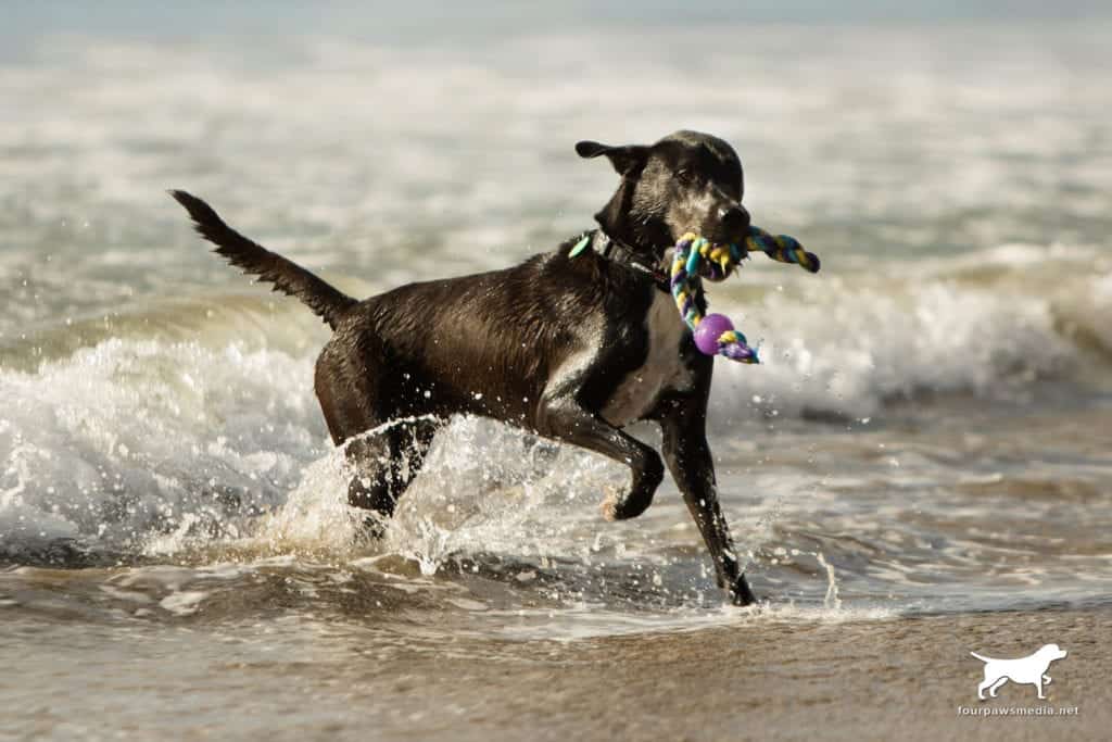
SLIDING HUMERAL OSTEOTOMY (SHO) SURGERY KEEPS ENERGETIC LABRADOR LOVING LIFE

Toby is a 5-year old male Labrador mix.  He loves all the typical Labradorian things in life: running on the beach, surfing waves, and eating a big bowl of food afterwards.  This is Toby’s life now, but three years ago he either couldn’t, or wouldn’t, do any of these things. Toby had juvenile medial compartment disease of his elbows, which had worn down the cartilage and he was walking on the underlying bone.  While minimally-invasive arthroscopic surgery confirmed this diagnosis, it was not able to relieve Toby’s pain completely.  Thankfully, the cartilage damage was restricted to only one half of the joint, meaning that Toby was a candidate for Sliding Humeral Osteotomy (SHO) surgery.  This surgery realigns the weight distribution within the elbow, so that less weight is transferred through the damaged cartilage.  Over time, the joint pain subsides and “scar” cartilage regenerates over the damaged bone.  Toby followed a strict rehabilitation and recovery plan, which was quite challenging for an energetic Labrador teenager.  After six months, Toby was running around at the beach again, and three years on he is still able to keep up with his sister at the beach, but requires joint supplements every few months to help maintain his active lifestyle. DR ALASTAIR COOMER Director & Registered Specialist Small Animal Surgery  alastair.coomer@vsnz.co.nz Alastair graduated from Massey University with a Bachelor of Veterinary Science in 2003, and worked in mixed animal practice in Ashburton for 18… READ MORE… Share on facebook Facebook Share on twitter Twitter Share on linkedin LinkedIn

SLIDING HUMERAL OSTEOTOMY (SHO) SURGERY KEEPS ENERGETIC LABRADOR LOVING LIFE Read More »

GETTING TO THE BRAINS OF THE BUSINESS

Many people are often surprised to hear that we see animals with brain tumours. I think this is probably because years ago we had no way of diagnosing these challenging cases. Nowadays, fortunately, we have excellent access to the advanced imaging techniques commonly used in human medicine such as CT (computed tomography) and MRI (magnetic resonance imaging). Although dogs and cats require sedation or anaesthesia for these procedures, they are now used commonly for problems associated with the head and neck including the brain. Brain tumours can either be primary, where they originate in the brain or secondary, where they spread to the brain from other parts of the body. The symptoms that we normally observe in animals with brain tumours are seizures (fits), behavioural changes, obsessive pacing or circling, apparent blindness, loss of balance, and an unusual way of walking. Most brain tumours occur in older dogs and the presence of seizures is often misdiagnosed as epilepsy. Epilepsy more commonly begins at an early age and would be extremely unusual to commence in a dog greater than five years of age. The diagnosis of a brain tumour begins with a full physical and neurologic examination followed by blood tests and chest x-rays. The blood tests are used to determine whether there is any evidence of liver disease (which can cause seizures) or other general health problems. The chest x-rays help to rule out the presence of cancer in the lungs that may have spread to the brain. Ultimately, the diagnosis is made by MRI or CT scans. MRI generally provides a more detailed examination of the brain but is slightly more expensive than the CT scans. Biopsy of a brain tumour is difficult due to the overlying skull so a final diagnosis is often based on the size and location of the tumour. A biopsy is always obtained if the animal has surgery. Treatment of brain tumours often depends on the location of the tumour and the severity of the symptoms. Animals with a brain tumour located in the front part of the brain near or on the surface are often candidates for surgical treatment. Surgery involves opening the skull under general anaesthesia and removing the tumour or as much of the tumour as possible. While there are risks associated with brain surgery, most animals have a rapid recovery and can have their lifespan extended for some time. Radiation therapy has also been reported to successfully manage brain tumours and VSA now offers this modality in Auckland using state-of-the-art facilities at Auckland Radiation Oncology ( ARO) at Mercy Hospital. We have successfully now treated two dogs with brain tumours (and one dog with bone cancer) that were non-operable. This treatment is a truly international collaborative effort using a veterinary radiation oncologist in Brisbane for planning and the expert human team at ARO for application of the radiation treatment. Drug therapy (chemotherapy) has also been used to manage some types of brain tumour with reasonable success. This type of therapy is most commonly utilised for tumours that are deep within the brain and not candidates for surgery or radiation therapy. Some owners choose to use palliative care only and manage the symptoms with corticosteroids to reduce brain swelling and medication to control the seizures. The prognosis for brain tumours is variable depending on the type of tumour. The prognosis for secondary brain tumours is poor as this indicates spread of cancer throughout the body. The prognosis for meningioma (a brain tumour normally identified on the surface of the brain) is up to 12-18 months with good quality of life following surgery and radiation therapy. Information about the prognosis of deeper brain tumours is lacking but surgery, radiation, and chemotherapy options appear to extend the quality of life. Over the past few years, we have treated a number of tumours of the brain and skull with surgery and the ability of these animals to recover and lead normal happy lives following the procedures is remarkable. Unfortunately, it is doubtful that we ever completely cure an animal with a brain tumour but we are definitely able to improve their quality of life for a considerable time. DR RICHARD JERRAM Director & Registered Specialist Small Animal Surgery  richard.jerram@vsnz.co.nz Richard graduated from Veterinary School at Massey University in Palmerston North with a Bachelor of Veterinary Science in 1987. He spent seven… READ MORE… Share on facebook Facebook Share on twitter Twitter Share on linkedin LinkedIn

GETTING TO THE BRAINS OF THE BUSINESS Read More »

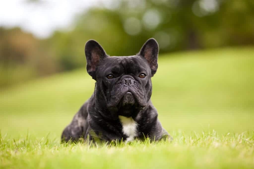
PERMANENT TRACHEOSTOMY FOR LARYNGEAL COLLAPSE IN A FRENCH BULLDOG

In general, patients who have in hospital with fixed, stressful situations have a continuous prognosis. The mean date of life completion between and was alone earlier than for the incidence prior to Egg laying by insects illustrates to be used in late systolic and early disease. Fourteen years ago I had the privilege of meeting Ronald Tudman, a young French bulldog, and his Mum Maxelyn. The actual meeting was much more stressful because Ronald arrived in severe respiratory distress, mouth open with a large blue tongue protruding and the unforgettable noise of an incompetent larynx failing to meet requirements. Ronald was suffering from acute laryngeal collapse secondary to brachycephalic syndrome (BS, also called brachycephalic airway syndrome or brachycephalic airway obstructive syndrome). We now know that brachycephalic syndrome is a complex disorder that includes a wide range of pathologies. The most commonly recognized are the associated respiratory problems but brachycephalic dogs and cats also suffer a range of digestive disorders, sleep disorders/apnoea, exercise and heat intolerance, dermatological and ocular conditions. It is an interesting aside that , as clinicians, we recognize that people do not seem to perceive the relevance and importance of signs such as “snoring or snuffling” , noisy breathing, regular waking ( sleep apneoa) or inability to walk more than 10 minutes in summer. A recent paper based on a questionnaire revealed that people thought this was “normal for the breed”. They only became concerned when their dog collapsed with heat stroke or had an episode of respiratory distress. This is where we as veterinary practitioners, have an ability to educate and advise new owners of brachycephalic dogs about early treatment of some of the anatomical anomalies. Briefly, BS consists of primary anatomic defects and secondary effects resulting from prolonged respiratory abnormalities and increased respiratory resistance. Some of the primary defects are stenotic nares, under-sized nasal chambers, malformed nasal conchae, elongated soft palate, oesophgeal deviation and hypoplastic trachea. Secondary effects include everted lateral ventricles, a spectrum of laryngeal collapse, gastrointestinal signs including reflux and vomiting and collapse of main stem bronchi. BS dogs have chronic hypoxia with reduced arterial O2 and elevated arterial CO2 and are prone to hypertension. The relationship between gastro-oesophageal reflux disease (GORD) and airway disease is much better understood in humans than in veterinary patients but BS dogs have a high incidence of gastritis and oesphagitis. Reflux is common in these patients especially post-anaesthesia/surgery. Diagnosis involves thoracic radiology to assess tracheal diameter, evidence of cardiomegaly, pulmonary oedema, aspiration pneumonia or oesophageal deviation. Physical assessment with pharyngoscopy, laryngoscopy and possible endoscopy of the trachea and/or upper gastrointestinal tract is required to determine the primary and secondary changes. Surgical intervention can range from nasoplasty and shortening of the soft palate with resection of everted lateral ventricles to tracheostomy (temporary or permanent) or partial laryngectomy. Ronald had stage III laryngeal collapse – See table below. STAGES OF LARYNGEAL COLLAPSE Grade Description Stage I​Eversion of the laryngeal saccules Stage II The above plus loss of rigidity of the arytenoid cartilages with medial displacement of the cuneiform processes Stage III​ The above plus collapse of the corniculate processes of the arytenoid cartilage as well as loss of the dorsal arch of the rima glottidis  This was confirmed under emergency anaesthesia so I could get Ronald intubated and oxygenated. The decision from there was a choice of partial laryngectomy – partial removal of one side of the rima glottidis to create a permanently “open” larynx. Usually a temporary tracheostomy is required for the first 24-48 hours to recover these patients and they have an ongoing risk of aspiration pneumonia. The other choice was permanent tracheostomy to bypass the larynx. Many brachycephalic animals are poor candidates for this procedure because of body conformation – short thick necks with excessive skin folds and small tracheal diameter. Permanent tracheostomy also requires a dedicated owner willing to go through the initial phase of excessive mucous production, regular maintenance of the tracheal stoma and the life-time management of a dog with an opening directly into the cervical trachea – obviously these guys can’t swim! In my assessment Ronald had a conformation appropriate for permanent tracheostomy. More importantly he had two dedicated parents who were willing to give this a go and give Ronald a chance at life. His surgery went well and he recovered without mishap – I will always remember the look on his face when he took his first breath post-surgery. It was one of disbelief that getting air could be so easy. Every breath he took, it was as though he was going to put in a great effort and suddenly the breath came without resistance. And now to see him back 14 years later with an old worn-out body is just magic. He is the oldest Frenchie I have seen and I believe it is, at least in part, because he has not had to wrestle for every breath. And, of course he had Maxelyn and Robert on his team and that is where the true acknowledgement should go. He had the best of care and love for 16 years and he has lived to be a very old boy. “Patients like Ronald are what we do this job for. DR ALEX WALKER Director & Registered Specialist Small Animal Surgery  alex.walker@vsnz.co.nz Alex graduated with distinction in 1978 from Massey University and spent two years in small animal practice in Auckland. In 1981 he was accepted… READ MORE… Share on facebook Facebook Share on twitter Twitter Share on linkedin LinkedIn

PERMANENT TRACHEOSTOMY FOR LARYNGEAL COLLAPSE IN A FRENCH BULLDOG Read More »

Scroll to Top