- All Posts
- Cases
- Pet Health
- VSA News

When Lottie arrived at VSA’s Sylvia Park clinic for spinal surgery, nobody could have predicted that the hardest part of...

At just one year old, Scout, a bright-eyed Miniature Dachshund with a soft spot for chicken, was facing a condition that, left...
If your pet has been found to have a heart murmur, an irregular heartbeat, or is showing signs that may...

Applications are now open for the Zoetis Rotating Internship at Veterinary Specialists Aotearoa. Run in partnership with Zoetis (formerly Pfizer)...

If your pet seems slower than usual, reluctant to jump or climb stairs, or just not quite themselves, pain could...

Hip scoring, dysplasia and osteoarthritis are terms that you may come across frequently when thinking about getting a new puppy,...

Ectopic Ureters – Marty’s rare condition Marty, an adorable golden retriever puppy, was just 15 weeks old when he first...
Parvovirus – what every dog owner should know We generally see an increase in cases of parvovirus over the summer...
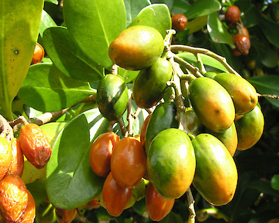
Did you know that Karaka Berries are highly toxic for dogs? The New Zealand native evergreen Karaka tree (Corynocarpus laevigatus)...
From spinal cyst to surgery: How Lottie found her way
When Lottie arrived at VSA’s Sylvia Park clinic for spinal surgery, nobody could have predicted that the hardest part of the day would be convincing her to go home again. Lottie, a dog with a rare spinal cyst causing progressive loss of function, had been referred to VSA specialist surgeon Dr Richard Jerram after her owner Alice Gasnier noticed something wasn’t right. Alice, a veterinary nurse at Companion Vets in Hamilton, knew enough about neurology to understand the road ahead wouldn’t be simple. “Lottie is a bit of an unusual case,” Alice said. “Richard noted she was a classic nurse’s dog, with a very unusual condition.” Cystic conditions of the tail end of the spinal cord are very uncommon in dogs but the more frequent use of advanced imaging studies with CT or MRI has improved our ability to diagnose this and to provide more focused treatment options” said Dr Jerram. The journey began with a consultation at the regional clinic service offered in Hamilton, a service that Alice said made an immediate difference for a dog who doesn’t travel well. “Lottie is not a big fan of travelling, so having the ability to get Richard’s expert opinion without the big car trip was a big plus,” she said. Dr Jerram walked Alice through the diagnosis and gave her an honest picture of what surgery could and couldn’t achieve. “Any spinal surgery is a big deal for both the dog and the owner. There are some considerable risks associated with the delicate nature of nerves and spinal cord tissue. These procedures require specialised surgical instrumentation, advanced lighting and intra-operative magnification. While these challenges and risks are present, VSA has a strong reputation and high success rate with these patients. Alice was able to much a thoroughly informed decision on whether she wanted to proceed with the surgery,” explained Dr Jerram. When the decision was made to go ahead, Alice and Lottie made the trip north to Sylvia Park. Alice said the clinic’s calm environment helped ease what could have been a stressful day, and that the team made both of them feel welcome from the moment they arrived. Updates came through that evening once Lottie was in recovery, and nurse Akiko called daily in the days that followed. “I looked forward to my daily updates on how Lottie was getting on,” Alice said. “I know she had been well cared for.” The proof came at discharge. When Lottie was brought into the room to go home, she turned around and walked straight back to the nurses! A few weeks later, she was back to see surgeon Dr Jerram for her six-week post-operative check. Richard was very pleased to see the impressive degree of progress in Lottie. Her neurologic function had improved dramatically and she was a very happy dog wagging her tail far better than before the procedure. From here, recovery is a team effort. Lottie is now working through an ongoing rehabilitation program with the VSA physiotherapy team, who have tailored a plan to rebuild her strength, balance, and confidence. For Alice, the experience was shaped as much by the communication as the clinical care, with updates shared between VSA and Lottie’s referring clinic throughout. “We are grateful to have access to such a skillful, caring team,” she said. “To be able to give our own pets — and our clients’ pets — the best possible outcomes.” About our regional service If you would like more information on this service, or to enquire on Dr Jerram’s visit schedule in your region, please get in touch with our team on info@vsnz.co.nz or call on 09 320 5645. Read more news Click Here Read more cases Click Here Our services Find out more
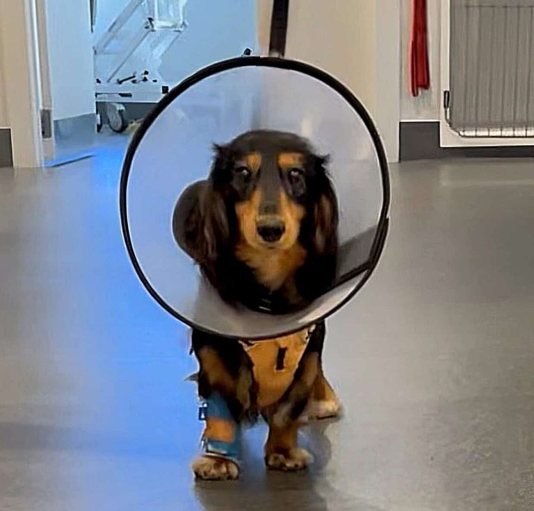
Scout’s Heart Journey
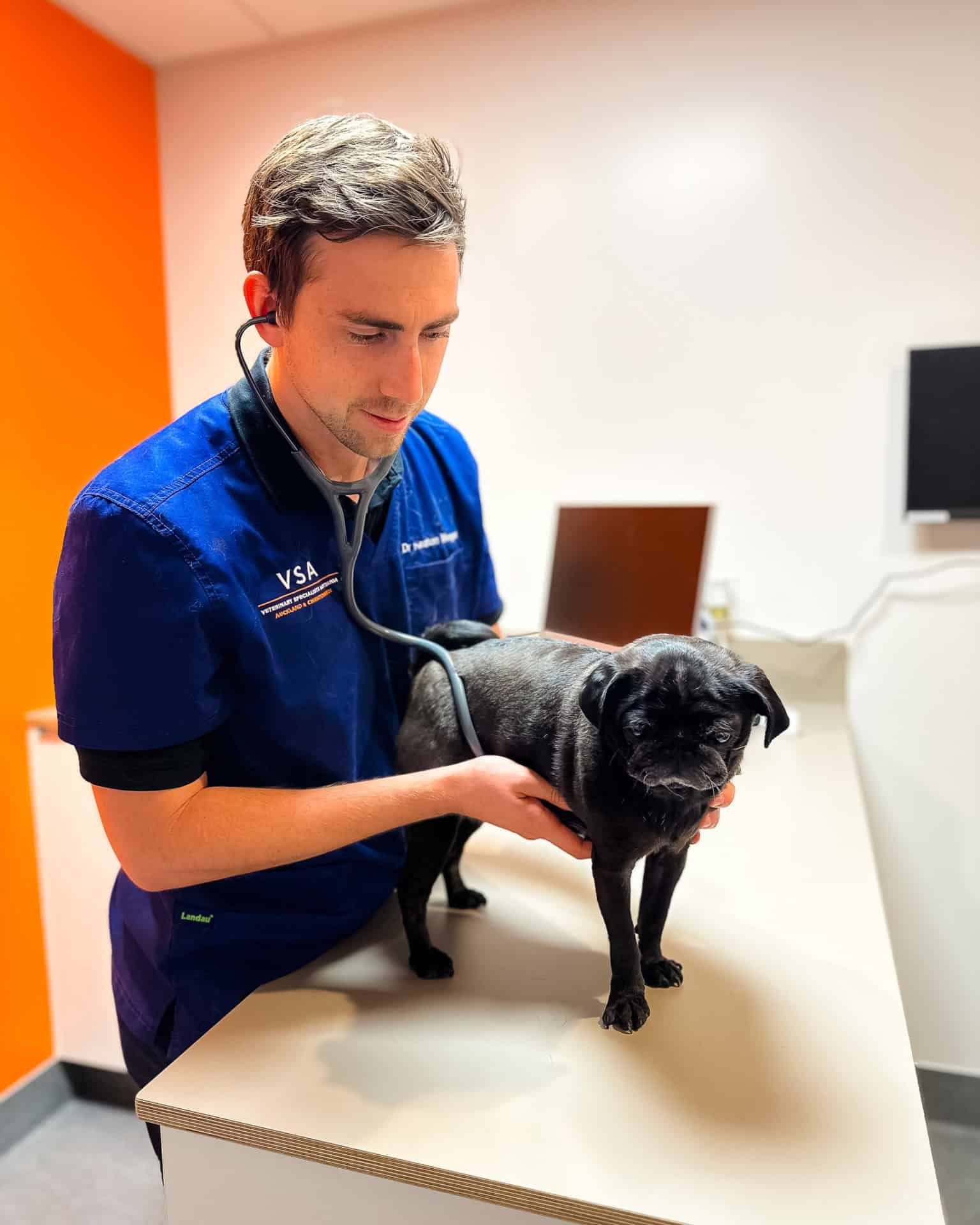
At just one year old, Scout, a bright-eyed Miniature Dachshund with a soft spot for chicken, was facing a condition that, left untreated, would have cut his life dramatically short. After a minimally invasive procedure with our cardiology team, he’s back home, running, snuggling, and looking for snacks like nothing ever happened. A heart problem from day one Scout was diagnosed with a patent ductus arteriosus, or PDA which is a congenital defect where a small blood vessel that should naturally close shortly after birth stays open. The result is abnormal blood flow between the heart’s two main arteries, putting extra strain on the heart muscle and, over the first couple of years of life, leading to heart failure. In Scout’s case, the strain was already taking its toll. Echocardiography showed marked enlargement of his left atrium and ventricle, early signs that his heart was working much harder than it should. Without intervention, his prognosis was poor. A precise, minimally invasive fix Under the care of cardiologist Dr Keaton Morgan, Scout underwent transcatheter PDA occlusion, a procedure performed through a small access point in his femoral artery, with no open-chest surgery required. A specialised device called an Amplatz Canine Duct Occluder (ACDO) was guided into position using live fluoroscopic imaging and placed inside the abnormal vessel to seal it shut. The result was the best one possible: complete closure of the PDA with no residual blood flow, confirmed on follow-up echocardiography. Scout’s heart beginning to return to a more normal size was already apparent with early signs of reverse remodelling. The minimally invasive method for placing an ACDO device is the most rewarding cardiac surgery I perform. The small incision in the leg is barely noticed by the patient and the rapid, complete occlusion (blocking) once the device is placed is satisfying for everyone; dog and doctor alike! Scouts rapid recovery was great to see and is the expectation for this procedure. Dr Morgan https://www.vsahospital.co.nz/wp-content/uploads/2026/06/PDA_5.mp4 Home with a healthier heart Scout recovered beautifully, with plenty of cuddles (and chicken) from the nursing team along the way. He’s now back home with an owner who, not long ago, was bracing for very different news. A case like Scout’s depends on tight teamwork, cardiology, imaging, anaesthesia, and nursing all playing their part. It’s also a reminder of what advanced interventional cardiology can do: turning a condition that would once have been life-limiting into the beginning of a long, happy dog life. We were recommended to take Scout up to Massey Vet Services, where they did all the checks and then gave us the options. To invest the time and money for Scout’s benefit was a no‑brainer for us, as he is such an important part of our family. From the initial phone conversation with the specialist at VSA, to meeting the team and seeing how welcoming they were, and then the care they showed for Scout, plus the follow‑up post‑operation call to put me at ease, to picking him back up when he was discharged… I could just see the love he was surrounded by, from people who genuinely cared for him. When I picked him up, he was like a new dog, with blood flowing freely again. He is a well‑loved member of our family. A massive thanks to the whole VSA team. – Marty, Scout’s owner Getting a referral To access our cardiology service, speak to your local vet, who can refer you to VSA. For more information, visit the cardiology services page or contact the team on 09 320 5645. Read more news Click Here Read more cases Click Here Our services Find out more

Specialist Cardiology Service Now Available at VSA
If your pet has been found to have a heart murmur, an irregular heartbeat, or is showing signs that may point to heart disease, such as reduced energy, coughing, difficulty breathing, or fainting, your local vet may refer you to a veterinary cardiologist for further assessment. Heart conditions in pets can range from manageable with the right medication to complex enough to require specialist intervention, and early diagnosis can make a significant difference. If you’ve noticed changes in your pet’s movement, behaviour, or mood, speak to your local vet about whether a referral to a pain specialist might help. About our cardiology service We offer a dedicated cardiology service led by specialist cardiologist Dr Keaton Morgan. The service investigates and treats a wide range of cardiac conditions, including mitral valve disease, heart murmurs, arrhythmias, congenital disorders, and all forms of heart disease. Initial assessment typically includes an echocardiogram — an ultrasound of the heart — and an ECG to evaluate the structure and function of the heart. Where needed, more advanced diagnostics are also available, including Holter monitoring, loop recorder implantation, CT scanning, and angiography. Outpatient echocardiograms are now available at VSA’s Auckland West hospital. This allows your pet to be assessed faster as an outpatient. Results are shared by your local vet and reviewed by our team, so a diagnosis or care plan can be put in place sooner. This makes specialist cardiac assessment more readily accessible for stable pets who may otherwise face a longer wait to be seen. Many of the pets I see are referred after their vet has picked up a murmur during a routine check, vaccine appointment or when being evaluated for a separate illness. This is often before the owner has noticed any changes at home. This is the best position to be in, because it gives us time to assess the underlying condition and if indicated, start treatment to prolong the “no symptom” stage of heart disease. This can help avoid cardiac symptoms, avoid surgery later in the animal’s life or increase stability if general anaesthesia is required for routine procedures such as dental cleanings. Dr Morgan Meet Dr Keaton Morgan Dr Morgan completed his internship at the University of Guelph in Canada, followed by his cardiology residency at the University of Minnesota in the United States. He brings international specialist training and is highly experienced in interventional procedures for congenital cardiac diseases and pacemaker implantation. Dr Morgan has a particular interest in expanding the knowledge base around cardiovascular diseases in animals and helping owners understand the condition present. His primary goal with every case is to maintain a great quality of life regardless of the disease present, and provide all options available to achieve this goal. Puppies and kittens Some puppies and kittens are born with heart conditions that show no obvious signs in their first weeks of life but without detection and treatment, these conditions can become life-threatening within months or first years of life. VSA offers an affordable echocardiogram for puppies and kittens at $450, making it easier for families to get peace of mind early. If the scan does identify something that needs attention, our cardiology team will guide you and your local vet through the options. Some congenital heart conditions are covered by pet insurance, so it is always worth checking your policy before your appointment. Additional therapies such as hydrotherapy, physiotherapy, and laser treatment may be recommended as part of your pet’s plan and can be arranged through VSA’s wider specialist team. Getting a referral To access our cardiology service, speak to your local vet, who can refer you to VSA. For more information, visit the cardiology services page or contact the team on 09 320 5645. Read more news Click Here Read more cases Click Here Our services Find out more

Applications open: Zoetis Rotating Internship 2027
Applications are now open for the Zoetis Rotating Internship at Veterinary Specialists Aotearoa. Run in partnership with Zoetis (formerly Pfizer) since 2001, the program is one of New Zealand’s most established pathways into specialist veterinary medicine. Of the 35-plus interns who’ve completed it, twelve have gone on to earn specialist status across surgery, radiology, internal medicine, cardiology, and dermatology. “Coming back to VSA as a veterinary cardiologist feels like coming full circle. It’s incredibly rewarding to work alongside the people who supported my early development, and to now contribute as a specialist in an environment that continues to value learning, collaboration, and patient-centred care.” Dr Keaton Morgan, Veterinary Cardiologist A year of rotations across our Auckland hospitals The 2027 intake begins in January, and over 12 months interns rotate through surgery, internal medicine, emergency medicine, cardiology, neurology, anaesthesia, and diagnostic imaging, working alongside specialists at our Auckland West and Sylvia Park hospitals. It’s a hands-on, immersive year. Interns shadow and assist with referral consultations, procedures and surgeries, and build real-world consulting confidence within our emergency service, always with experienced support close by. What it’s like to be part of our team VSA is New Zealand’s leading specialist veterinary organisation, operating three world-class specialist and emergency hospitals nationwide. What makes the program stand out, year after year, is the team behind it and the opportunities that come from the experience. “Starting my career as an intern at VSA gave me an exceptional foundation — not just clinically, but in how I approach complex cases and work as part of a specialist team.” Dr Isobel MacEwan, Radiology Resident We are looking forward to welcoming vets ready to take their first step into specialist practice. Applications close Sunday 21 June 2026 Apply now Questions? Please contact Recruitment Manager, Bridget Cassidy, at careers@vsnz.co.nz. Read more news Click Here Read more cases Click Here Our services Find out more

Helping pets live more comfortably: VSA’s pain management clinic
If your pet seems slower than usual, reluctant to jump or climb stairs, or just not quite themselves, pain could be the reason. Chronic pain in pets often has a gradual onset and is easy to miss as animals are instinctively good at hiding discomfort. What looks like ‘slowing down with age’ can sometimes be a manageable condition with the right care. If you’ve noticed changes in your pet’s movement, behaviour, or mood, speak to your local vet about whether a referral to a pain specialist might help. About our pain management clinic VSA’s Pain Management Clinic was started by Dr Yasmine Messiaen to give pets with chronic pain access to specialist assessment and a treatment plan tailored to their individual needs. The clinic takes a comprehensive approach, combining advanced clinical expertise with genuine care for both the pet and their family. Meet Dr Yasmine Messiaen Dr Messiaen is a Board-Certified Small Animal Surgery Specialist and a Diplomate of the American College of Veterinary Surgeons, the highest level of recognition in her field, achieved in 2025. She completed her veterinary degree at St. George’s University, followed by internships and a surgical residency in the United States and Canada. Her expertise spans minimally invasive surgical techniques and chronic pain management, and she founded VSA’s Pain Clinic with a clear purpose: to help pets live their happiest, most comfortable lives. Outside of work, Yasmine can be found on outdoor adventures with her partner, or tending to her quirky chickens. What to expect A pain clinic appointment begins with a thorough physical examination, including an assessment of your pet’s mobility, overall health, and pain levels. Dr Messiaen will discuss your goals and concerns for your pet, allowing her to develop a tailored treatment plan that supports their specific needs, lifestyle, and quality of life. Additional therapies such as hydrotherapy, physiotherapy, and laser treatment may be recommended as part of your pet’s plan and can be arranged through VSA’s wider specialist team. Introductory offer for new clients To book into the pain clinic at VSA Auckland West, speak with your local vet to get a referral. To make it easier for families to access specialist pain care, VSA is currently offering an introductory package of $550, which includes initial consultation and one-month recheck, for new pain clinic clients. Regular price $620. Medications and additional therapies not included. Read more news Click Here Read more cases Click Here Our services Find out more

Hip Dysplasia: What is it and what can you do about it?
Hip scoring, dysplasia and osteoarthritis are terms that you may come across frequently when thinking about getting a new puppy, researching new breeds or simply wondering why your own dog is a little bit slower to get up or has an unusual hindlimb gait. Hip dysplasia is the abnormal development of the coxofemoral (hip) joints. It is a condition seen mostly in larger dog breeds, most commonly German Shepherds, Golden Retrievers and Labradors. However, the condition can occur in any breed and infrequently occurs in cats too. Hip Dysplasia is not immediately evident at birth. This syndrome is developmental and progresses with age. There is a significant genetic component to hip dysplasia hence the importance of hip scoring and selective breeding. There are however other influences such as environmental influences. A good example of this would be over-feeding. Over-nutrition during a puppy’s first year of life, in which rapid growth of the bones and joints occurs, is thought to contribute to the development of dysplastic hips. The pathophysiology of hip dysplasia includes multiple variables and the disease can be unilateral or bilateral and range in severity. The coxofemoral joint can be thought of as the ball (the femoral head) and the socket (the acetabulum) joint (see figure 1). Figure 1: This radiograph has been annotated to show the “ball” of the femoral head and the “socket” of the acetabulum. The primary abnormality in these dogs is poor congruency of the coxofemoral joint (figure 2), in other words the ball does not sit in place as it should. This can lead to instability and the abnormal exertion of forces on the cartilaginous linings of the joint. Figure 2: This patient has severe incongruency and laxity of the coxofemoral joints. The earliest changes noted include the erosion of the cartilage on the femoral head and acetabulum, joint effusion where fluid builds up in the joint and thickening of the ligament on the femoral head. These changes cannot be detected with X-rays, however, clinical signs might include a swaying hindlimb gait, reduced range of motion in the hip joints and reluctance to climb stairs. As the disease progresses, the body attempts to stabilise the lax joint by producing new bone around the femoral head, neck and acetabulum (figure 3). The bone underlying the damaged cartilage hardens, a process known as sclerosis. As more new bone is deposited around the joint, the femoral neck becomes thickened and the head loses its rounded shape. These degenerative changes are collectively called ‘osteoarthritis” and are the cause of pain and reduces mobility. Unfortunately, there is no method of reversing osteoarthritic changes once they have developed. Treatment options include certain surgical procedures which either remove, alter, or replace the painful, dysplastic joint, or medical therapy to reduce pain and improve mobility. Due to the heritable component, ‘hip scoring’ of higher risk breeds prior to breeding is important. This involved potential breeding animals having a set of radiographs (X-rays – PennHIP) taken by a certified veterinarian or veterinary technician where the hips are forced into both compressed and distracted positions. The radiographs are sent to the USA to measure the degree of laxity of the hips and determine if there are already signs of osteoarthritis. A numerical value called the ‘distraction index’ is then assigned to each dog. Not only does the distraction index quantitively measure the laxity in each joint, it has been shown to correlate with the risk of developing osteoarthritis in the future. This makes in an excellent test for young dogs with PennHip being able to be performed as early as 16 weeks of age. Hip Dysplasia is a debilitating disease that can significantly limit an animal’s quality of life. Ethical and responsible breeding with testing of potential parents, appropriate early nutrition and limited high intensity activity during growth are all important factors in reducing the incidence of hip dysplasia. If you are considering a new puppy, have questions about hip scoring or are worried about the hip health of your existing furry friend, consultation with your veterinarian is highly recommended.