Cases Archives - Veterinary Specialists Aotearoa | Veterinary Specialist and Animal Referral Services in NZ with 3 specialist animal hospitals in Auckland and Christchurch

Cases

Dedicated Neurology Department Available at VSA

VSA offers a dedicated neurology service, led by Dr Sam Khan, BVetMed, PhD, MRCVS. Sam graduated from the Royal Veterinary College in the United Kingdom and completed his internship, PhD, and neurology residency at the University of Cambridge. His research background includes spinal cord injury, intervertebral disc disease, and immune mediated neurological disorders. Neurological conditions can be complex and difficult to diagnose, and assessment by a neurologist can make a significant difference to outcomes. Neurological conditions can present with a wide range of clinical signs including pain, weakness, seizures, altered behaviors and movement abnormalities and the close integration of this service across our specialist departments enables us to support the most complex neurological cases. I strongly believe in patient-led, individualised care. Every patient and family is different, so I enjoy working with owners to understand what matters most to them and making decisions together about the best approach for their pet. I’m proud to be part of Veterinary Specialists Aotearoa and really value working alongside such a knowledgeable, supportive and compassionate team who always strive for the best outcome for our patients. Dr Sam Khan Advanced Electrodiagnostics VSA has a new electrodiagnostic system enabling faster more accurate assessment of neuromuscular conditions. This advanced technology supports nerve conduction studies, electromyography (EMG), and evoked potential testing, providing our specialists with valuable diagnostic information to assess nerve and muscle function. This enhanced capability allows VSA to deliver a more comprehensive and efficient diagnostic service for patients with suspected or confirmed neuromuscular disease. Case study – Freddie Freddie presented to our emergency team following a number of collapse-like episodes which were precipitated by excitement. He was subsequently referred to our Neurology Department, where he was assessed by our neurologist, Dr Sam Khan. Following a thorough neurological examination and careful observation of Freddie’s episodes, Dr Khan was able to recognise a pattern consistent with narcolepsy. Narcolepsy is a sleep disorder that can cause muscle weakness or collapse, often triggered by strong emotions such as excitement. Although it can look very alarming, the episodes are usually brief, and patients quickly return to normal. A comprehensive range of investigations including bloods, liver function tests, infectious disease tests, CT and spinal tap was undertaken to exclude an underlying cause of Freddie’s episodes. All of Freddie’s results were within normal limits, helping to rule out a number of potential underlying causes. Freddie was subsequently started on imipramine. Since beginning the treatment, Freddie has since done very well, allowing him to get back to enjoying life. Freddie’s case highlights the importance of careful observation and thorough investigation when presented with unusual collapse-like episodes and demonstrates how recognising the less typical signs of neurological disorders can help lead to an accurate diagnosis and appropriate treatment. Getting a referral To access our neurology service, contact your local vet, who can refer you to VSA. For more information, visit the neurology services page or contact the team on 09 320 5645. Read more news Click Here Read more cases Click Here Our services Find out more

Dedicated Neurology Department Available at VSA Read More »

How Bella and Chi found relief at VSA’s pain management clinic

Bella and Chi’s owners knew something wasn’t quite right. Although their dogs were showing signs of discomfort, the cause was not immediately clear. Despite their dedication to improving their wellbeing, it took time to recognise that chronic pain was playing a significant role in the changes they were seeing. Bella: from growls to cuddles Bella, a white Bearded Collie cross adopted from the council, was born around September 2012. She spent most of her life as an active, ball-obsessed dog. About three to four years ago, her family noticed she was slowing down. Then, over the last year or two, her personality seemed to change too. She began giving a low growl whenever people or the other dogs in the house got too close. Her family put it down to age. It turned out to be pain. Since starting the right pain medication with Dr Yasmine at VSA Auckland West, in Henderson, Bella is herself again. She still enjoys the slower pace of an older dog, shorter walks and plenty of naps, but she’s back to cuddling and playing with toys, even rolling over the family board game mid-play just to get a pat. Her family describes the change as incredible, and they’re grateful for the care and attention she’s received. The signs of chronic pain in dogs and cats can be subtle. Owners frequently describe their pets are being more irritable, refusing to be touched, slowing down, or simply not being themselves. Changes in behaviour can appear gradually over time and are often dismissed as the pet “just getting old”. However, these changes may be signs of discomfort says Dr Yasmine. “At the VSA Pain Clinic, each pet is carefully assessed to find the source of pain and underlying cause so a tailored plan can be developed. Our goal is to help pets feel more comfortable and enjoy a better quality of life. If your pet’s behaviour or mobility has changed, don’t assume it’s simply old age. Recognising signs of pain is the first step towards helping them.” Dr Yasmine  Messiaen, Registered Specialist Small Animal Surgery. Chi: a rough start, a remarkable recovery Chi, a brindle Staffordshire Bull Terrier cross Dachshund adopted from the Henderson Animal Shelter, was born around January 2014. Her unusual body shape, a Dachshund’s length combined with a Staffy’s solid build, has always been hard on her spine. At around four years old, she was paralysed from the neck down. She made a remarkable recovery after hyperbaric chamber treatment, learning to walk again, and even developed a range of different barks to communicate during that time, a habit she still has today. Despite everything, Chi has always been a friendly, sociable dog who stops to greet everyone she meets. Working with Dr Yasmine, her family has come to realise that Chi has likely been dealing with some level of pain or discomfort for most of her life. With the right medication now in place, she’s able to jump on and off the couch again and even join the school run on foot, with a stroller on standby for when she gets tired. The common thread For both pets, the biggest change has been the same: they are more themselves, calmer, less reactive, and more able to enjoy everyday life. “Bella and Chi are both such a big part of our family and they were quietly living with pain before we realised. Now that they’re on the right medication, they’re calmer, happier and so much more themselves. Dr Yasmine is so great with the dogs and explaining the process to us and where we can expect to see improvement. We wish we’d known about the pain clinic sooner, but we’re so grateful we found our way there.” Pri, Bella and Chi’s pet mum and owner. Bella and Chi’s stories are a good reminder that behaviour changes in senior or long-term unwell pets, like withdrawal, irritability, or reduced activity, aren’t always just “getting older.” They can be signs of manageable pain. About our pain management clinic VSA’s Pain Management Clinic was started by Dr Yasmine Messiaen to give pets with chronic pain access to specialist assessment and a treatment plan tailored to their individual needs. The clinic takes a comprehensive approach, combining advanced clinical expertise with genuine care for both the pet and their family. Read more news Click Here Read more cases Click Here Our services Find out more

How Bella and Chi found relief at VSA’s pain management clinic Read More »

From spinal cyst to surgery: How Lottie found her way

When Lottie arrived at VSA’s Sylvia Park clinic for spinal surgery, nobody could have predicted that the hardest part of the day would be convincing her to go home again. Lottie, a dog with a rare spinal cyst causing progressive loss of function, had been referred to VSA specialist surgeon Dr Richard Jerram after her owner Alice Gasnier noticed something wasn’t right. Alice, a veterinary nurse at Companion Vets in Hamilton, knew enough about neurology to understand the road ahead wouldn’t be simple. “Lottie is a bit of an unusual case,” Alice said. “Richard noted she was a classic nurse’s dog, with a very unusual condition.” Cystic conditions of the tail end of the spinal cord are very uncommon in dogs but the more frequent use of advanced imaging studies with CT or MRI has improved our ability to diagnose this and to provide more focused treatment options” said Dr Jerram. The journey began with a consultation at the regional clinic service offered in Hamilton, a service that Alice said made an immediate difference for a dog who doesn’t travel well. “Lottie is not a big fan of travelling, so having the ability to get Richard’s expert opinion without the big car trip was a big plus,” she said. Dr Jerram walked Alice through the diagnosis and gave her an honest picture of what surgery could and couldn’t achieve. “Any spinal surgery is a big deal for both the dog and the owner. There are some considerable risks associated with the delicate nature of nerves and spinal cord tissue. These procedures require specialised surgical instrumentation, advanced lighting and intra-operative magnification. While these challenges and risks are present, VSA has a strong reputation and high success rate with these patients. Alice was able to much a thoroughly informed decision on whether she wanted to proceed with the surgery,” explained Dr Jerram. When the decision was made to go ahead, Alice and Lottie made the trip north to Sylvia Park. Alice said the clinic’s calm environment helped ease what could have been a stressful day, and that the team made both of them feel welcome from the moment they arrived. Updates came through that evening once Lottie was in recovery, and nurse Akiko called daily in the days that followed. “I looked forward to my daily updates on how Lottie was getting on,” Alice said. “I know she had been well cared for.” The proof came at discharge. When Lottie was brought into the room to go home, she turned around and walked straight back to the nurses! A few weeks later, she was back to see surgeon Dr Jerram for her six-week post-operative check. Richard was very pleased to see the impressive degree of progress in Lottie. Her neurologic function had improved dramatically and she was a very happy dog wagging her tail far better than before the procedure. From here, recovery is a team effort. Lottie is now working through an ongoing rehabilitation program with the VSA physiotherapy team, who have tailored a plan to rebuild her strength, balance, and confidence. For Alice, the experience was shaped as much by the communication as the clinical care, with updates shared between VSA and Lottie’s referring clinic throughout. “We are grateful to have access to such a skillful, caring team,” she said. “To be able to give our own pets — and our clients’ pets — the best possible outcomes.” About our regional service If you would like more information on this service, or to enquire on Dr Jerram’s visit schedule in your region, please get in touch with our team on info@vsnz.co.nz or call on 09 320 5645. Read more news Click Here Read more cases Click Here Our services Find out more

From spinal cyst to surgery: How Lottie found her way Read More »

Marty, a gorgeous golden retriever puppy with ectopic ureters

Ectopic Ureters – Marty’s rare condition

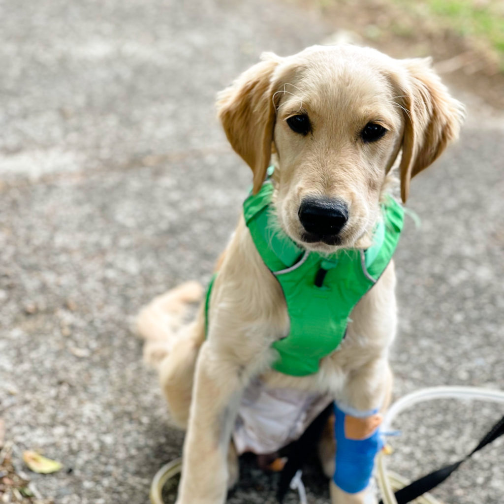
Ectopic Ureters – Marty’s rare condition Marty, an adorable golden retriever puppy, was just 15 weeks old when he first presented to the VSA team in Sylvia Park. He presented initially to the medicine department for urinary incontinence which was occurring when Marty was awake, lying at rest and when asleep. The owners adopted Marty at 8 weeks old and first noticed signs of urine leakage approximately one week later. Marty was otherwise a bright and clinically well puppy.  Further investigation was needed to determine the underlying cause of Marty’s incontinence. Marty was kept in hospital for a day as Dr Wen-Jie Yang recommended a collection of diagnostic tests that initially consisted of blood work (biochemistry and haematology panel), a urine sample for culture, urinalysis, and an abdominal ultrasound.  The biochemistry renal parameters of creatinine were within normal reference range with phosphate mildly elevated. Urine culture, urinalysis and haematology were all unremarkable. Abdominal ultrasound showed that the kidneys were severely enlarged (>95mm length) with marked hydronephrosis (swelling of the kidneys).  Both ureters (tubes that transport urine from the kidneys to the bladder) were severely distended (>33mm diameter) with a highly tortuous path and terminated at the cranial aspect of the prostate gland. Conclusions of the ultrasound showed that Marty had bilateral ureteral ectopia with very severe secondary hydroureter and hydronephrosis caused by ureterocoeles. A ureterocoele is simply a swelling limited to the end of the ureter as it enters the bladder and can be associated with urine refluxing backward to the kidney causing enlargement due to a build-up of urine (hydronephrosis). The ureterocoeles were likely responsible for both Marty’s incontinence and hydronephrosis. Ectopic ureters (abnormal location of the ureters) are a rare finding in dogs but are the most common congenital anomaly that causes urinary incontinence. The disease is thought to arise because of disruption in nor­mal embryogenesis (the process of embryo development). Hydronephrosis leads to kidney damage, however, Marty’s most recent kidney parameters had improved compared to previous blood testing which was good news. While Marty was not displaying obvious pain, hydronephrosis can be a painful disease and he was sent home with gabapentin to help control any underlying pain.  The severity of this condition meant that without surgery, Marty would soon succumb to end stage renal failure with a grave prognosis. It became clear from the diagnostic test results that Marty was a puppy with a big problem. Surgery was Marty’s last hope, and he was transferred to the surgical team at VSA. A CT scan of Marty’s abdomen with a negative contrast cystography (an injectable contrast dye to help visualise the bladder) was recommended to further characterise the urinary tract pathology and facilitate pre-surgical planning.  This scan was performed a week later. CT scans are an excellent form of imaging as they offer a much higher level of detail compared to standard X-rays and can create a 360-degree view of the body’s internal structures. Dr Richard Jerram reviewed the CT results and discussed Marty’s significant urinary tract abnormalities and surgical options with his owners. Results confirmed bilateral extramural ectopic ureters. The extreme nature of the bilateral hydroureter and hydronephrosis was indicative of a chronic partial obstruction at the point where the ureters enter the bladder (ureteral termination) whilst the severe distension of the urinary bladder was thought to be due to compression of the bladder neck by the overlying ureters. Surgical treatment was scheduled a week later and consisted of abdominal surgery to perform neo-ureterostomy bilaterally. This procedure involves creating a new ureter opening where the distal portion of the ureters are relocated so that they enter the bladder in the correct anatomical location.  Even though this surgical approach would aid in resolving Marty’s immediate problem (relieving urine build up) and hence improve renal function, Marty’s urinary incontinence was expected to continue. Further surgery may be necessary, assuming that Marty does not progress into renal failure. This would involve placement of ureteral stent devices (subcutaneous ureteral bypass device and a hydraulic urethral sphincter occlusion device) to improve the incontinence by maintaining the flow of urine from the kidneys to the bladder when the ureter becomes obstructed.  Marty, being a resilient little puppy, recovered well from major urinary tract surgery.  An indwelling urinary catheter was placed to assess his urine flow. He appeared to be producing good urine from the kidneys with no evidence of further renal deterioration indicating that the neo-ureterostomy sites were flowing well.  Once it was clear that Marty could urinate without the need for the urinary catheter, he was discharged 3 days post-surgery with pain relief, antibiotics and strict 2 week confinement – no easy task for a energetic puppy!  One month following his ectopic ureter surgery, Marty has been making excellent progress at home. He is now able to urinate with no evidence of incontinence. A repeat abdominal ultrasound evaluation was performed which confirmed functioning ureters with some renal improvement but with continued dilation of the ureters bilaterally.  While Marty is not out of the clear yet, with the help of his committed owners, he still has a long road to recovery that involves repeat ultrasound exams and bloodwork to assess his ongoing renal function. Due to the severe nature of Marty’s condition, his long-term prognosis remains uncertain.  The VSA team have our fingers crossed for Marty and he looks like he is heading in the right direction – go Marty!

Ectopic Ureters – Marty’s rare condition Read More »

A gorgeous German Shepherd puppy is seated on the ground. He is one of Police Dog Vali's newest litter

Auckland Police’s Newest Recruits

Happy news for Vali the Police Dog Here at VSA we are lucky enough to work with some incredible animal services throughout New Zealand, including Auckland Zoo and the NZ Police Dog Section. The hard-working police dogs and their handlers are well known to our team and so we were incredibly proud to be able to help out police dog Vali with her second pregnancy. Vali is a 3-year-old German Shepherd who works for the Auckland Police Dog Unit. She came in to see our Mount Albert team at the end of September for a pregnancy ultrasound to check for a positive pregnancy. On this visit, Dr Mike Coleman confirmed multiple viable gestational sacs, but we were unable to say how many there were.  It’s tricky to do this when there are multiple sacs at the best of times, let alone when you are doing an ultrasound on a very active police dog. Vali is a very strong dog, as all police dogs are and so getting her to stay still while we tried to count the number of pups was a challenging task. Vali is a dog who knows her own mind and that meant that while on this visit we were able to confirm a pregnancy, we had to defer counting how many puppies there might be to another visit. Vali’s pregnancy continued incredibly well, and we had a second visit from her at the end of October. On this visit we took some x-rays and were able to confirm 7 puppies. You can see then numbered on the x-ray below.  Shortly after this second visit, Vali took a trip down to the Dog Training Centre birthing unit and she started whelping almost immediately. By the end of the weekend of 29/30th October she had delivered 7 healthy puppies, 4 girls and 3 boys.  Vali is an amazing mum. She is calm and maternal, using all her police dog training to keep her puppies in check. Female police dogs are made of tougher stuff than the males – well you would need to be to rear 7 new police dogs, wouldn’t you ?! Vali’s pups are growing up fast and they are just over 7 weeks old now. Aren’t they adorable?  Meet Auckland’s newest Dog Squad!

Auckland Police’s Newest Recruits Read More »

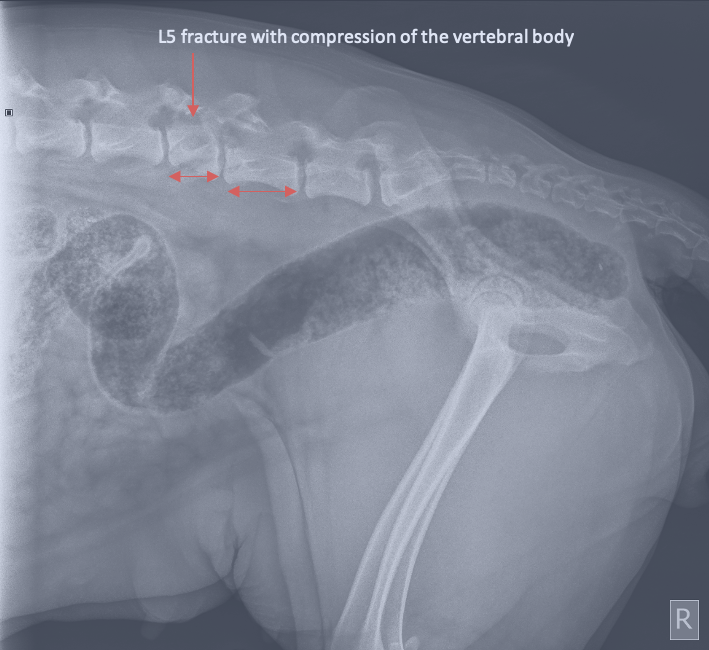
x-ray showing cassie's compression fracture

CASSIE, A NEW MUM WITH A FRACTURED SPINE

Cassie was only eighteen months old when she first came to see the VSA team in Christchurch. She had recently given birth to her first litter of puppies and was a proud new mum of nine little huskies. Three weeks after their birth, she was having a bit of a break and was playing outside when she was hit and rolled by a quad bike. She managed to walk back to her owners before collapsing and was then rushed to the after-hours vet clinic. When she arrived, Cassie was in pain and unable to use her back legs, although she could still wag her tail at people! She was started on intravenous fluids and strong pain relief. Radiographs (X-rays) of her spine were taken which showed a compression fracture of the 5th vertebra in her lumbar spine (l5). Cassie was well looked after by the after hours team overnight and was transferred to VSA the next day for further evaluation and treatment. Initial radiography/x-ray showing a compression fracture of Cassie’s L5 vertebra When Cassie arrived at VSA she was still laterally incumbent and couldn’t walk. She had reduced reflexes in her left hindlimb which indicated some dysfunction to the nerves traveling to her left leg but luckily, she was still able to feel and move both back legs by herself. Dr Alastair Coomer recommended a CT scan of her spine as soon as possible to further evaluate the degree of trauma and make a plan on how to fix her spine. Meanwhile, Cassie’s nine puppies had to stay at home with their human family so that she could rest and recover quietly in hospital. A CT scan was performed the next day which confirmed that Cassie had multiple fractures through the pedicle, the lamina the articular facet and the body of her 5th lumbar vertebra. Being a more sensitive imaging modality, the CT also showed that the disc space between the 5th and 6th lumbar vertebrae was misaligned and there had been both ventral (downwards) and lateral (sideways, away from the midline) displacement of the caudal (back) part of Cassie’s vertebral column. CT Images of a normal L5 vertebra (left) and Cassie’s L5 vertebra (right) with multiple fractures Dr Coomer reviewed the CT images and the next day Cassie was taken to surgery to stabilise her spine. The fractured vertebra was identified and the spinal column was realigned back into its normal position. The spine was then stabilised using two metal ‘string of pearl’ (SOP) plates. These plates spanned three of her vertebrae which provided stability to her spine and were held securely in place by a total of 10 screws. The surgery was a success and Cassie spent the night in hospital recovering from her big procedure. post-operative radiographs showing Cassie’s fracture stabilisation with SOP plates and screws. With her new bionic back, Cassie was up and walking the very next day, with only a little support from a sling. She was able to go home and see all her puppies again, although she would need to be confined to a crate for the next 6 weeks to help with healing. Cassie became a regular at the veterinary physio, stretching and strengthening her muscles to help her back to tip-top shape.  Two and a half weeks after surgery, Cassie was doing incredibly well. She was able to stand by herself and walk for short periods before getting tired. She was building up the muscles in her legs and although she still had some reduced reflexes in her left hind left this was expected to continue improving with time. Unfortunately, Cassie re-presented to the after-hours clinic two days later as she had become suddenly painful and was not herself. She was diagnosed with Pyometra, a severe infection of her uterus, and underwent another surgery to perform an ovariohysterectomy (spey). Once again, she recovered well from the procedure and was discharged the next day. She was welcomed home by her babies and whilst Cassie won’t be having any more puppies in the future, these nine have kept her busy enough! Since her surgeries, Cassie has continued to make excellent progress. Recovering from spinal surgery for a dog is no mean feat. It takes a lot of work, a deteremined dog and very committed human parents. Cassie is one lucky lady to have all three and she is well on the way to a full recovery. She’s getting regular physio and hydrotherapy, has started going to longer walks and will be back to running around and being a typical husky in no time at all! Share on facebook Facebook Share on twitter Twitter Share on linkedin LinkedIn

CASSIE, A NEW MUM WITH A FRACTURED SPINE Read More »

Figure 2b Hemipelvectomy

THE ROLE OF HEMIPELVECTOMY IN DEFINITIVE AND PALLIATIVE TREATMENT OF PERIPELVIC NEOPLASIA: A CASE STUDY

GRACE KAEMPERBVScV student INTRODUCTION Hemipelvectomy is a surgical procedure involving removal of the hemipelvis and corresponding hindlimb.  It can be classified as total or subtotal, where subtotal has three further subtypes: mid-to-cranial, mid-to-caudal and caudal (Barbur et al. 2015). There is a paucity of veterinary literature detailing both technical and clinical aspects of hemipelvectomy.  One study presents hemipelvectomies as “complex, aggressive surgical procedures” with a high risk of intraoperative complications and “a slower return to ambulation than amputation” (Kramer et al. 2008).  There is a need to challenge this commonly held perception in the veterinary industry, and the purpose of this case report is to present hemipelvectomy as a viable treatment option for a range of otherwise inaccessible tumours. This case study examines three dogs that underwent hemipelvectomy in 2018 at a single veterinary specialist hospital in Auckland.  Two dogs presented with osteosarcoma of the proximal femur and ilium respectively, and the third was diagnosed with a peripheral nerve sheath tumour (PNST) of the sciatic nerve.  In all three cases, the location of the tumour rendered standard limb amputation (coxofemoral disarticulation) insufficient for primary tumour control.  Therefore, a hemipelvectomy was required to obtain appropriate surgical margins. All dogs were followed from diagnosis and surgery, through to recovery, to examine both the oncologic and functional outcomes.  See Table 1 for a summary of the three cases. HEMIPELVECTOMY PROCEDURE   Anaesthesia protocols were determined at the discretion of the surgeon (two surgeons performed three surgeries).  A similar anaesthesia protocol was used in all three cases.  Dogs were pre-medicated I/V with 3 µg/kg fentanyl (Fentanyl Citrate Injection 500µg; Hospira New Zealand Ltd, Auckland, NZ) or 3 µg/kg remifentanil (ULTIVA; Pharmacy Retailing Ltd, Auckland, NZ), which was followed by anaesthetic induction using I/V 0.25 mg/kg diazepam (Ilium Diazepam; Ethical Agents Ltd, Auckland, NZ) and propofol (Fresofol 1%; Fresenius Kabi, Auckland, NZ) to effect.  A constant rate I/V infusion (CRI) of lidocaine (Lopaine 2%, Ethical Agents Ltd, Auckland, NZ), ketamine (Ketaset 10%, Zoetis New Zealand Ltd, Auckland, NZ) and fentanyl/remifentanil (depending on the pre-medication) and I/V fluid therapy with lactated Ringer’s solution at 10 mL/kg were given for the duration of the procedure and recovery.  In two cases, 0.1 mg/kg morphine (DBL Morphine Sulphate 5 mg/mL Injection, Hospira) was given epidurally for additional analgesia.   The dogs were placed in lateral recumbency with a hanging leg preparation and an anal purse string suture was placed.  The surgical technique for all three cases was similar and based on the hemipelvectomy method described by Barbur et al. (2015).  A ventral approach was initially used with elevation of the sartorius, pectineus, gracilis, and adductor magnes et brevis muscles from their origin.  This allowed isolation, ligation, and transection of the femoral artery and vein, and the obturator nerve.  Pubic and ischial osteotomies were performed.  Further muscle elevations exposed the ventral aspect of the sacroiliac joint, which was then disarticulated.  The limb was then adducted, and the approach changed to dorsal.  The sciatic nerve was isolated and transected.  The ilium was freed by elevating its muscle and soft tissue attachments.  The sacrotuberous ligament and dorsal sacroiliac ligament were incised to free the ilium from the sacrum and to allow removal of the hemipelvis and ipsilateral limb.  The pelvic diaphragm was reconstructed during closure by approximating the remaining musculature.  The subcutaneous tissue and skin were closed routinely to minimise tension (Barbur et al. 2015).  A Jackson Pratt drain was placed before closure.  All resected tissues were submitted for histopathology.   Any differences peculiar to the individual cases are discussed below. CASE ONE A 13-year-old, female, spayed Labrador Retriever presented with acute left hindlimb lameness of 10 days’ duration.  Radiographs revealed a pathological fracture of the proximal left femur with suspected neoplastic involvement of the ipsilateral coxofemoral joint.  Thoracic radiographs showed a 5 mm-diameter, rounded, soft-tissue nodule in the caudodorsal aspect of the right caudal lung lobe.  Definitive tissue sampling of the pulmonary nodule was not obtained, and both benign and malignant considerations were given during clinical decision making.  An abdominal ultrasound identified a bilobed, diffusely heterogenous, soft-tissue mass of 31 mmx 12 mm effacing the left adrenal gland, and mild left medial iliac lymphadenopathy.  Preoperative biopsy of the fracture site was not pursued, as treatment was with palliative intent. A subtotal mid-to-cranial hemipelvectomy with concurrent amputation of the left pelvic limb was performed, utilising an osteotomy of the ilium caudal to the sacrum, osteotomy of the pubis within the obturator foramen, and osteotomy of the cranial ischium.  Soft tissue dissection was performed outside the muscular compartment containing the fracture, such that the fracture haematoma and tumour were not encountered during surgery.  Haemostasis was achieved with an electrothermal vessel sealing device (Ligasure; Medtronic Corporate ANZ, Auckland, NZ) and no complications arose during surgery. Fig.1 Dorsoventral radiograph of the pelvis of a Labrador Retriever taken 10 weeks after hemipelvectomy surgery as palliative treatment for osteosarcoma of the left proximal femur and acetabulum.  There is an aggressive monostotic lesion of the right ischium with motheaten lysis, heterogenous sclerosis and irregular periosteal new bone formation. Histopathology identified the proximal femoral/acetabular malignancy as an osteosarcoma with haemorrhage and necrosis.  Surgical margin assessment revealed no neoplastic cells within 40 mm of the inked margins. The dog was strongly ambulatory following surgery and was discharged from hospital the day following her hemipelvectomy.  The functional outcome, as reported by the owner, was excellent for 10 weeks after surgery.  At that time, the dog developed acute non-ambulatory paresis and urinary incontinence.  Neurologic examination was consistent with a painful L7-S3 lesion.  Radiographs revealed motheaten lysis and heterogeneous sclerosis of the right ischium, and further pulmonary nodules (Figure 1).  Advanced imaging and tissue biopsy were declined, but distant metastasis (spine and lungs) was suspected.  The dog was discharged with analgesia provided by 400 mg gabapentin (Neurontin; Pfizer New Zealand, Auckland, NZ) and 37.5 mg carprofen (Rimadyl; Zoetis New Zealand Ltd, Auckland, NZ) both given orally twice daily, but was euthanised due to lack of clinical improvement. CASE TWO An 11-year-old, male, neutered

THE ROLE OF HEMIPELVECTOMY IN DEFINITIVE AND PALLIATIVE TREATMENT OF PERIPELVIC NEOPLASIA: A CASE STUDY Read More »

MIRACLE MAGGIE'S MEDICAL MYSTERY: SUBCUTANEOUS URETERAL BYPASS

MIRACLE MAGGIE’S MEDICAL MYSTERY

When Maggie’s mum noticed she wasn’t eating and slept in longer than usual one Sunday morning, she knew something was wrong and brought her in to VS A&E. Maggie is no stranger to us at VSA and we know how much she loves food, so even though she looked fine on the outside, her history of dietary indiscretion, oesophageal foreign bodies, ureteral obstructions and chronic kidney disease had us wondering what was happening inside. Little did we know at the time she would become a very complex case.

MIRACLE MAGGIE’S MEDICAL MYSTERY Read More »

ELBOW DYSPLASIA IN DOGS

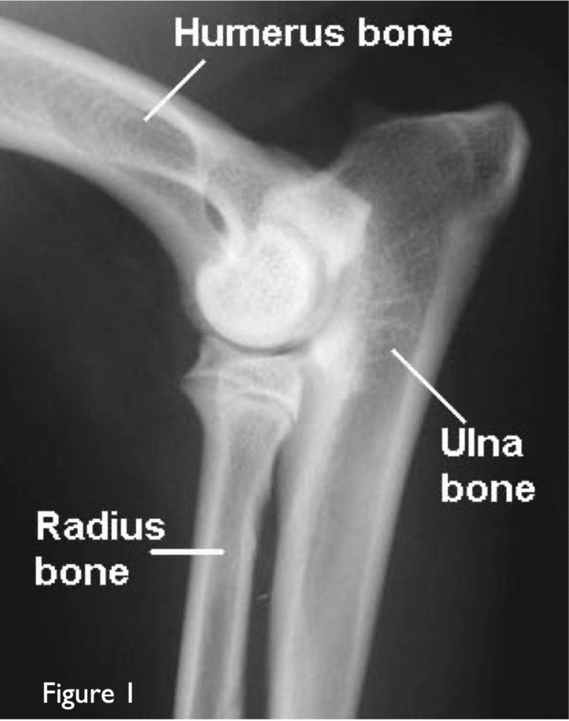
The most common cause of front leg lameness in the dog is developmental problems of the elbow joint. This disease leads to degenerative changes (osteoarthritis) in the joint including carti- lage damage, bone spur (osteophyte) pro- duction, and loose bone fragments. ELBOW JOINT FUNCTION The elbow joint of the dog is formed by three bones (the humerus, ra- dius and ulna bones {Figure 1}) that must fit together correctly for the joint to func- tion normally. There are ligaments on the inside and outside of the elbow to main- tain stability. Although the elbow joints of dogs and humans have similar anatomy, the forces applied to the dog’s elbow are different because dogs stand on their front legs. The humerus bone should glide smoothly around the C-shaped curve formed by the radius and ulna bones. CAUSES OF ELBOW DYSPLASIA Elbow dysplasia is typically a developmental, inherited disease of young large -breed dogs. In some dogs, during growth, a step develops in the C-shape between the radius and ulna bones possibly due to the radius bone being too short or too long relative to the ulna.  This causes extra stress to be placed on prominent parts of the ulna bone (the anconeal or coronoid processes {Figure 2}) or the cartilage of the humerus. Cracks can then occur in the bone across the lines of greatest stress or the bone fails to grow normally. The joint cartilage may also be damaged in this process. Arthritis develops as a result of the damage to the bone and the cartilage. There are three main forms of elbow dys- plasia that cause pain and arthritis: Fragmented coronoid process (FCP). A crack occurs in this triangular- shaped bony bulge on the inside edge of the ulna bone. Ununited anconeal process (UAP). A lack of normal bone growth re- sults in this bone fragment becoming loose at the top of the ulna bone. Osteochondritis of the humerus (OCD). A flap of loose cartilage develops on the inside surface of the humerus bone. SYMPTONS Typically, affected dogs show front leg lameness that can start as young as 5 months of age. The lameness may be in- termittent and tends to improve with rest and get worse with exercise. Not all dogs with elbow dysplasia show symptoms. DIAGNOSIS A thorough orthopaedic and neuro- logic examination is performed evaluating the dog when walking and by manipulating all four limbs and the spine. Swelling, pain, and joint stiffness can frequently be felt in the affected elbow. Sedation or anaesthe- sia and X-rays are necessary to show signs of arthritis and to assess the bone structure. A CT scan will often be more effective in showing any bony changes. MEDICAL TREATMENT Some dogs with confirmed elbow dysplasia will improve without surgery. The object of medical treatment is to control the symptoms. The dog should not be allowed to get overweight and ex- ercise should be controlled. Cartilage- protecting agents (omega fatty acids, glu- cosamine, green-lipped mussel, fish oil) may help lubricate the joint and keep cartilage healthy. Generally, lifelong supplementation is necessary. Anti-inflammatory medication (aspirin-like drugs) can be helpful in reducing pain but should only be necessary occasionally. Medical treatment does not stop arthritis from continuing to develop. SURGERY Other dogs with elbow dysplasia benefit from surgery depending on the type of damage and arthritis. The type of surgery differs from dog to dog and may involve passing a small telescope (arthroscope) into the joint to see if a step is present between the radius and ulna bones. Loose bone fragments may then be removed or stabilized using a bone screw. (Figure 3) The ulna bone may be cut to remove the step and a pin may be placed to help healing. (Figure 4) RESULTS Dogs that have had surgery generally show excellent results with some progression of arthritis, few complications and normal function. If the ulna bone is cut, healing takes about 2-3 months and most dogs can return to full activity 4-5 months after surgery. Be sure to check out our article on post operative care for dogs with elbow dysplasia for best results post operation.

ELBOW DYSPLASIA IN DOGS Read More »

PEBBLES’ BILATERAL HIP REPLACEMENT JOURNEY

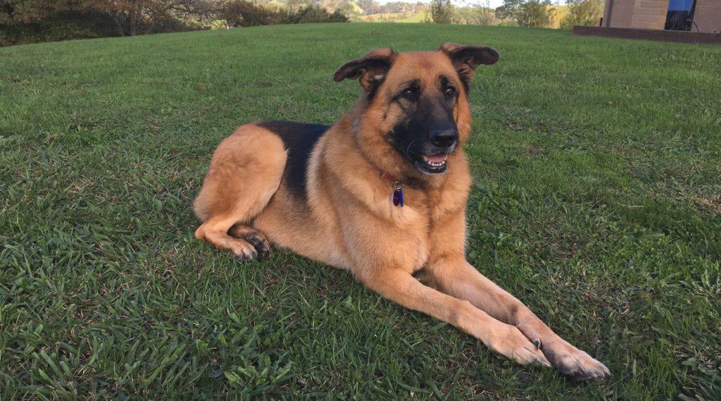
Pebbles was an active four-year-old German Shepard with a passion for chasing seagulls on the beach. Even though the seagulls would fly away before she caught them, her enthusiasm for her game never wavered. Over a period, Pebbles started to show less enthusiasm for her chase, often choosing to sit and watch instead. One day after a visit to the beach, her owners noticed her limping. Thinking it was because she had pulled something, they kept her quiet at home for a few days in the hope that it would clear up. After several days with no improvement, they took her to their local vet where she was prescribed anti-inflammatories. Pebbles showed no noticeable improvement on the medication, so she returned to the vet who referred her to us for further examination. Once here, we did some x-rays to determine what was happening. Her x-rays showed Pebbles had hip dysplasia in both hips, with her left being the worst. Once we talked with her owners, they said it explained a lot of her recent behaviour. She did not like going outside as much anymore, nor would she follow them in the garden. Instead, she returned to the house and slept. With uncontrolled pain restricting her quality of life, her owners knew she could not continue as she was. Fig.1 Initial radiographs/x-rays of Pebbles’ hips showing arthritis in both joints. To treat Pebbles, we had the options of prescribing anti-inflammatory medication for the rest of her life, which was not very successful at reducing her pain, commiting to long-term lifestyle adjustment or physiotherapy programme, or a hip surgery. Her family decided that a left hip replacement would be the best treatment for Pebbles, even though they were anxious about the risks and complications of hip replacement surgery. The surgery went fine and to help her recover, Pebbles was confined to a cage for the next 12 weeks. After a final check, Pebbles could resume her normal daily activities. Her owners noticed an almost instant change in their dog. She went from being a reclusive and depressed “older” girl to an enthusiastic adult puppy who once again enjoyed chasing her beloved seagulls. A few months later, her owners noticed she was slowing down again. Pebbles showed signs of being depressed and was this time limping on her right leg. They returned here and we took further x-rays of Pebbles. This time they showed her right hip needed a replacement, so her owners decided to have this done one year after her left hip was replaced. Once again, surgery was successful. Unfortunately, when Pebbles returned to have her sutures removed, she fractured her femur on her right leg while walking outside in the carpark. Miraculously, the fracture did not involve the prosthetic hip. We repaired the fracture with a plate and Pebbles recovered as predicted. Fig.2 & 3 Radiograph/x-ray showing both hip replacements, but also the break in the right femur. Hip replacement surgery does have a 20-30% risk of complications. Some of these complications are severe, such as a fractured femur which happened to Pebble. However most of the complications can be fixed and result in a normal active life for the animal afterwards. Pebbles’ life has changed dramatically since her surgeries. Instead of being left to a life of poorly managed pain and a young dog who would rather sleep than do anything else, they have a dog which shows obvious enjoyment of life. Pebbles runs, plays and chases seagulls as much as she ever did, in a life free from pain and regular medications. DR ALASTAIR COOMER Director & Registered Specialist Small Animal Surgery  alastair.coomer@vsnz.co.nz Alastair graduated from Massey University with a Bachelor of Veterinary Science in 2003, and worked in mixed animal practice in Ashburton for 18… READ MORE… Share on facebook Facebook Share on twitter Twitter Share on linkedin LinkedIn

PEBBLES’ BILATERAL HIP REPLACEMENT JOURNEY Read More »

Scroll to Top