Common Heart Conditions And Procedures
Mitral Valve Disease
Mitral valve disease is the most common heart condition we treat in dogs and cats. It is a progressive condition which occurs when the valve between the left atrium and ventricle becomes thickened and distorted over time.
As the valve’s structure changes, it no longer closes properly, allowing blood to leak backward through the valve. This leakage leads to an enlargement of the left atrium and creates increased pressure within the heart chambers. Eventually, this pressure can cause fluid to build up in the lungs, a condition known as pulmonary oedema.
We offer two groundbreaking treatments for dogs with this condition to extend both the quality an quantity of life; Left Atrial Decompression and Trans Catheter Edge to Edge Repair (TEER).
Patent ductus arteriosus (PDA) occlusion
A PDA is a birth defect in the heart caused by incomplete changes in the heart’s circulation when an animal is born. In most cases it is usually picked up in an initial puppy or kitten check-up where a heart murmur is heard.
The ductus arteriosus is an important blood vessel that ensures that blood does not go to the lungs unnecessarily as the fetus is developing in the uterus. During the first few hours after birth, this blood vessel would naturally close off. This allows blood to travel normally through the lungs for oxygenation as the lungs begin to function when the animal takes its first breath. In some baby animals, the ductus arteriosus remains open (patent). This results in serious, life-threatening changes in the way that the heart pumps blood through the heart and to the rest of the body.
Once a patient has been diagnosed with a PDA the goal of treatment is to close the open ductus arteriosus.
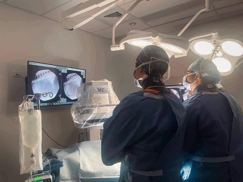
At VSA this can be accomplished through cardiac catheter-based (minimally-invasive surgery) occlusion. Catheter based occlusion is a minimally-invasive key-hole procedure and the patients usually go home the following day.
Pulmonic stenosis balloon valvuloplasty
Pulmonic stenosis is a congenital heart defect of the valve that is found between the right ventricle and the pulmonary artery. It is most commonly seen in breeds such as Labradors, Samoyeds, Terriers and brachycephalics.
At VSA we use a technique called balloon valvuloplasty in the treatment for valvular pulmonic stenosis.
This treatment involves the introducing of catheters via the veins and a deflated balloon placed through the abdominal stenotic valve. When the balloon is inflated, it opens the restricted valve leaflets. Measurements of the pressure gradient before and after treatment give information as to the success of the treatment.
Artificial pacemaker placement
An artificial pacemaker is placed in dogs with dangerously slow heart rates because of damage or aging of the conductive tissue.
The artificial pacemaker is able to take control of the heart rate to prevent it getting too slow, or stopping altogether. The pacemaker can also recognise movement to increase the heart rate with exercise. The result is prevention of death from slow/ stopping hearts and improved quality of life.
Here at VSA we have access to the latest pacemaker technology, allowing the pacemakers function and battery life to be monitored remotely.
Cutting balloon procedures
Subaortic stenosis is a narrowing of the area underneath the aortic valve, that causes an obstruction or blockage of the blood flow through the heart. The narrowing can be classified as mild, moderate, or severe. In moderate or severe cases the heart is forced to work harder and potentially be harmful to the heart’s health.
A cutting balloon is an inflatable vascular balloon with micro blades attached. The balloon is carefully positioned across stubborn obstructions through key hole surgery into the peripheral vessels. This type of balloon can be used to open more restrictive obstructions such as severe subaortic stenosis. Subaortic stenosis is a fibrous-muscular narrowing under the aortic valve which in particular cases may respond to a cutting balloon procedure.
Intravascular stenting
Stents are metallic expandable tubes that can be carefully implanted across tough, stubborn obstructions that respond poorly to a balloon inflation. The stent keeps the area open, eliminating the obstruction. This can be used for a range of vascular obstructions within the cardiovascular system, By opening up the region permanently, bloodflow can move freely and not back-up around the body.
Intravascular coiling
Coils are small metallic structures covered in a fine feather -like material. The feathers promote clot formation. Therefore, coils can be used to block abnormal vascular connections and can be delivered in a minimally -invasive procedure through a vessel in the neck or leg.
The most common procedure that requires the use of vascular coils in intra-hepatic portosystemic shunts, however at VSA they can also be used to occlude other abnormal vessels if deemed necessary.
If you think your pet requires specialist cardiology treatment, please contact your GP vet in the first instance.