LARYNGEAL PARALYSIS
For treatment of breathing difficulties in older dogs
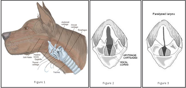
LARYNGEAL FUNCTION
SYMPTOMS
PROGNOSIS
DIAGNOSIS

MEDICAL TREATMENT
SURGERY
COMPLICATIONS
RESULTS


DISK FUNCTION
The intervertebral disk of the dog acts as a cushion between the spinal bones (vertebrae) to absorb the shocks and movements of normal activity. The normal disk is like a “jelly doughnut” with a gelatinous centre and an outer ring of stronger fibrous tissue. In certain breeds of dog (Dachshund, Poodle, Beagle, Spaniel, Corgi), the disk degenerates at a very early age. As the dog ages, the jelly like component of the disk becomes more gritty and less resistant to pressure. The disk is then no longer able to cushion the spine and the contents of the centre may forcibly squirt out and bruise the spinal cord. Alternatively, the outer part of the disk may bulge up putting pressure on the spinal cord.
DIAGNOSIS
A thorough neurologic examination is performed evaluating the head, all four limbs, and the spine. Pain can frequently be felt at the site of the affected disk. Anaesthesia and X-rays can help to show signs of narrowed disk spaces and degenerative disks. To confirm the diagnosis, a CT scan is recommended as this provides us with the best information on the spine. A special X-ray test called a myelogram can be helpful, in addition to a CT scan. Some dogs have an associated instability of the vertebrae that has contributed to the disk degenerating.
SURGERY
Dogs that have severe neck pain.or significant spinal cord damage are also candidates for surgery. The most common procedure is a ventral slot, which involves drilling a slot in the base of the vertebrae to relieve the spinal cord pressure and allow the delicate extraction of the disk material. Sometimes, the central portion of adjacent degenerative disks is removed to reduce the chance of further disk
SYMPTOMS
Syleptoms typically develop, in order of severity, from neck pain to weakness and wobbliness then finally to unwillingness to stand depending on the speed or the amount of the disk rupture. In the most severe cases, dogs lose the ability to walk and become quadriplegic.
MEDICAL TREATMENT
Some dogs with only mild symptoms will respond to medical treatment. Generally this involves three or four weeks of strict confinement to a cage with the dog only allowed out to go to the toilet. Pain relief (cortisone or aspirin-like medication) is given at the same time but this does not mean the dog can be more active. Dogs that do not improve or get worse with medical treatment are candidates for surgery.
RESULTS
Dogs that have retained the ability to walk prior to surgery have a 95% chance of complete recovery following surgery. Complete healing of the spinal cord can take up to six months to occur. Regular progress in spinal cord recovery is seen during this time. Dogs with unstable vertebrae have an increased risk of further instability developing later in life.
Rupture
For dogs with instability of the vertebrae, additional surgical stabilisation may be performed. This is done using metal plates and screws or bone cement.
PROGNOSIS
The prognosis for recovery is mostly dependent on the severity of the damage to the spinal cord. The ability to walk before treatment is the key indicator for prognosis in dogs with a slipped disk in the neck.
POSTOPERATIVE CARE
EXERCISE CONTROL
To allow the larynx to heal following the surgery, complete restriction of exercise is necessary for the first 2-3 weeks. Your dog can be walked on a lead for toileting. Light (5-15 minutes) lead walks can begin after 2 weeks.
BANDAGE AND SUTURE REMOVAL
A bandage is generally placed over the operated site for protection. This bandage should be removed 4-5 days after surgery. The skin stitches need to be removed 10-14 days following surgery. These tasks can be done by your regular veterinarian. Please call our hospital if there is any swelling, discharge or redness around the stitches.
FEEDING MANAGEMENT
As the larynx area will be swollen after surgery making swallowing difficult, it is very important that your dog is not allowed to eat watery or crumbly food after surgery. It is best to make up small meat-balls and feed your dog by hand each meatball for the first 2-4 weeks after surgery. Water intake also needs to be carefully monitored so that your dog is not gulping in large amounts of water at one time.
MEDICATION
Most dogs are sent home with medication for additional pain relief. Sometimes, antibiotics are also dispensed. Give the medications as prescribed. Further pain relief can be prescribed if necessary. Please let us know two days before suture removal if you think more medication is required or you may be charged an urgent fee.
BREATHING MANAGEMENT
Some dogs will cough and gag a small amount after surgery which is normal. Warm, humid environments should be avoided. Sometimes, excessive barking can occur putting a strain on the internal stitches, behavioral control can help this but anti-anxiety medication may be prescribed. You may notice a change in your dog’s voice after surgery, this is normal and occurs even when surgery is not performed.
Monitor your dog for signs of lethargy, decreased appetite and a moist cough, this could indicate pneumonia. If this occurs then an appointment with our hospital or your regular veterinarian is strongly recommended.
